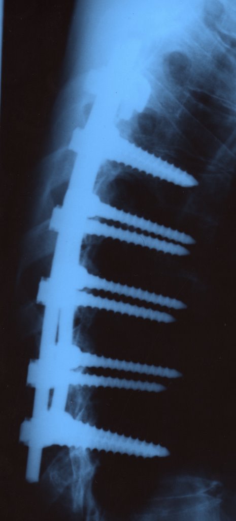
Thoracic Spinal Fusion
two screws in each vertebrae.
This feels very familiar.
Chronic pain often presents sufferers with a real "catch 22" dilemma. If they talk about their pain, they risk being perceived and labeled as hypochondriacs, or even worse?fakers or malingerers. On the other hand, if they hide their pain, others don't believe the pain is significant. It is enough to tax the patience of the most stoic person....
Compression and Wedge FracturesSpineuniverse.com
Thomas A. Zdeblick, M.D.
Professor and Chairman Orthopaedic Surgery
University of Wisconsin
Madison, WI, USA
What is a Compression/Wedge Fracture?
A compression fracture is a common fracture of the spine. It implies that the vertebral body has suffered a crush or wedging injury. The vertebral body is the block of bone that makes up the spinal column.Each vertebral body is separated from the other with a disc. When an external force is applied to the spine, such as from a fall or carrying of a sudden heavy weight, the forces may exceed the ability of the bone within the vertebral body to support the load. This may cause the front part of the vertebral body to crush forming a wedge shape. This is known as a compression fracture. If the entire vertebral body breaks, this is considered a burst fracture and is discussed elsewhere. The compression fracture may range from mild to severe in terms of severity. A mild compression fracture causes minimal pain, minimal deformity and is often treated with time and activity
modification.
Severe Pain
A severe compression fracture may be such that the spinal cord or nerve roots are involved, as they are draped over the sudden angulation of the spine. This may cause severe pain, a hunched forward deformity (kyphosis) and rarely neurologic deficit from spinal cord compression.
Risks - Osteoporosis - Trauma
The risk for spinal compression fracture increases with age. Osteoporosis is the most common risk facture for compression fractures. Osteoporosis is a condition in which there is thinning of the bones, weakening them. This may be due to a lack of calcium in the diet, certain medications, old age, inactivity or genetic factors. In general, some trauma occurs with each compression fracture. In cases of severe osteoporosis, the trauma may be minimal, such as, stepping out of a bathtub or lifting a heavy object. Moderate trauma is usually required to create a fracture in patients with mild to moderated osteoporosis. This may range from falling off a chair to an automobile accident. A normal spine may also suffer from a compression fracture when there is a severe forward bending injury. This most commonly occurs from a fall from a height or an automobile accident.
Nerve Injury
Neurologic injury is rare with compression fractures. The degree of neurologic injury is usually due to the amount of force present at the time of injury. If there is severe angulation of the spine secondary to a wedge fracture, this may stretch the spinal cord and create injury. This would then lead to loss of strength and sensation, as well as reflexes. In most patients with osteoporotic compression fractures, there is no neurologic injury but only pain from the fracture. However, if left untreated the fracture angulation may worsen and lead to late paralogic injury.
Diagnosis
A compression fracture is usually diagnosed by the history, physical exam and x-rays. In any patient over the age of 60 with the acute onset of sudden low back pain, a compression fracture should be suspected. Physical exam will usually note tenderness directly over the area of pain as well as mild kyphotic deformity (e.g., a sudden angulation forward or hunched over appearance). Plain x-rays will demonstrate the wedge shape of the vertebral body on a lateral view. A CAT scan is occasionally needed to help differentiate a compression fracture from a burst fracture.Occasionally an MRI scan is obtained to rule out disc herniation along with a compression fracture. MRI scan may also help differentiate pathologic compression fractures, that is, those that involve a tumor, from a typical osteoporotic compression fracture. In any patient with a known history of cancer, a compression fracture should tip off the physician to look for evidence of a metastatic lesion and pathologic fracture. If osteoporosis is suspected, a Bone Mineral Density (BMD) test may be ordered. This test helps determine the severity of the bone thinning. In addition, laboratory tests to look at blood count and thyroid function may be indicated as well. A decision as to whether to treat osteoporosis should be made by the
patients' primary physician.
Burst Fractures:Defined and DiagnosedSpineuniverse.com
Thomas A. Zdeblick, M.D.
Professor and Chairman Orthopaedic Surgery
University of Wisconsin
Madison, WI, USA
Thomas A. Zdeblick, M.D.
Professor and Chairman Orthopaedic Surgery
University of Wisconsin
Madison, WI, USA
What is a Burst Fracture?
A burst fracture is a descriptive term for an injury to the spine in which the vertebral body is severely compressed. They typically occur from severe trauma, such as a motor vehicle accident or a fall from a height. With a great deal of force vertically onto the spine, a vertebra may be crushed.If it is only crushed in the front part of the spine, it becomes wedge shaped and is called a compression fracture. However, if the vertebral body is crushed in all directions it is called a burst fracture. The term burst implies that the margins of the vertebral body spread out in all directions. This is a much more severe injury than a compression fracture for two reasons. With the bony margins spreading out in all directions the spinal cord is liable to be injured. The bony fragment that is spread out toward the spinal cord can bruise the spinal cord causing paralysis or partial neurologic injury.
Also, by crushing the entire margin of the vertebral body the spine is much less stable than a compression fracture.
Nerve Injury
Neurologic injury from a burst fracture ranges from no injury at all to complete paralysis. The degree of neurologic injury is usually due to the amount of force that is present at the time of the injury and the amount of compromise of the spinal canal. With a greater amount of force, more bony fragments can be forced into the spinal canal causing greater loss of spinal cord function. This may cause loss of strength, sensation or reflexes below the level of the injury.Typically, in a burst fracture that occurs at the junction of the thoracic and lumbar spines paralysis of the legs and loss of control of the bowel and bladder may result. In an incomplete spinal cord injury only partial paralysis or reflex loss is seen. With mild burst fractures only transient symptoms may be present or no neurologic injury may be present.
Severe Pain
Burst fractures cause severe pain. Typically, this is pain at the level of the fracture, that is, in the back. However, pain may also be present in the legs following the distribution of the affected nerves. Many patients complain of an electric shock type sensation into their legs when there is spinal cord compression. Most patients with a burst fracture are unable to walk immediately after the injury. Seldom, the patient may walk away from an accident and still have a burst fracture. However, often the amount of pain that is present is severe enough that patients know it is a good idea not to walk.
Diagnosis
At the scene of the accident, patients complaining of severe back pain should not be placed into a seated for flexed position. They should be kept lying flat and transported in the flat position. A patient who stands or sits with a burst fracture may increase their neurologic injury. Burst fractures require immediate medical care by an orthopedic or neurosurgeon trained in spinal surgery. The patient should be transported to an emergency room and x-rays obtained.The diagnosis of a burst fracture is usually made by x-rays and a CAT scan. Occasionally, an MRI scan may be ordered as well, in order to assess the amount of soft tissue trauma, bleeding or ligament disruption. The review of the CAT scan and x-rays allows the treating physician to make a determination as to the level of the fracture, whether it is a compression fracture, burst fracture or fracture dislocation, and to determine the amount of spinal canal compromise and spinal angulation. All of these factors enter into the treatment decision process.
The physical exam should be performed to document both spinal deformity, that is, angulation of the spine or tenderness of the spine at the level of fracture, as well as, a neurologic exam.
Neurologic exam should include testing of the muscle strength, sensation and reflexes of the lower extremities, as well as, testing of bowel and bladder sphincter control.
No comments:
Post a Comment